The Eurographics Association’s workgroup on Visual Computing in Biology and Medicine organises a biennial competition to acknowledge the contribution that computer graphics and visualization are playing in medicine and the life sciences, and to encourage further development in the field.
Originally called the Eurographics Medical Prize, the competition was renamed the Dirk Bartz Prize for Visual Computing in Medicine in 2010, in honour of Dirk Bartz, who passed away far too early in March 2010. Dirk Bartz was a highly recognised and enthusiastic scientist, teacher and promoter of visual computing in medicine, an active member of the Eurographics Association, and chair of the EG Medical Prize in 2007 and 2009. In 2021 the competition was renamed once more to the Dirk Bartz Prize for Visual Computing in Medicine and Life Sciences, to also include advances in biology and the life sciences.
Until 2019 the prize was collocated with the Eurographics Conference. From 2021 onwards it is collocated with EG/VGTC EuroVis. Details about how to apply are announced in the conference calls in odd-numbered years.
Hall of Fame
All winning entries, with links to the full papers in the Eurographics Digital Library.
2025 2023 2021 2019 2017 2015 2013 2011 2009 2007 2005 2003
2025
EuroVis 2025, Luxembourg City, Luxembourg, 2–6 June 2025
First Prize: Visual Disease Stories: Empowering Health Literacy and Promotion
Abstract
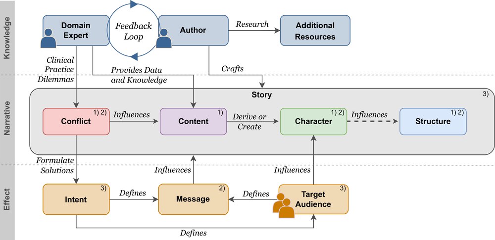
Narrative medical visualization bridges the gap between limited health literacy and the need for health communication. We explore disease stories to promote preventive behaviors, educate patients about their conditions and treatment options to foster informed consent, which is embraced by clinicians for its potential to alleviate the burden on healthcare systems and providers. We integrate data visualization with storytelling to make scientific information more accessible to diverse audiences. However, the expansive design space presents significant challenges, as design choices can greatly influence levels of engagement, memorability, and comprehension. Our work investigates the fundamental dimensions of the narrative design space – conflict, content, character, and structure – with the aim of maximizing its impact on disease communication.
Second Prize: Too Heart to Handle? Exploring Self-Directed And Collaborative Virtual Learning Environments in Anatomy Education
Abstract
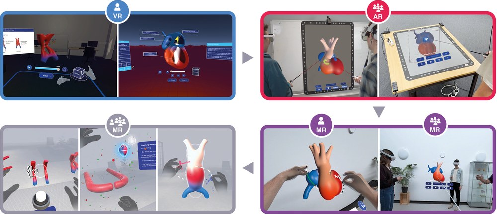
The integration of Extended Reality (XR) into medical education represents a transformative shift, particularly in anatomy training, where immersive simulations enhance cognitive engagement and knowledge retention. The developing heart is characterized by rapid morphological changes within a short time frame, which poses a significant pedagogical challenge. Conventional 2D imaging and static models often fail to convey these processes, limiting learners’ ability to conceptualize critical spatial relationships – a barrier in understanding congenital anomalies. To address these limitations, this work leverages XR-driven visualization and interaction paradigms to create virtual learning environments. Based on this, we propose methods for designing XR educational modules that adapt to both collaborative and self-directed learning contexts, using embryonic cardiogenesis as an illustrating case study. We present findings from mixed-methods user studies involving a total of 264 students, along with feedback from lecturers, highlighting the importance of an iterative, user-centered design approach.
Third Prize: X-ray simulations with gVirtualXray in medicine and life sciences
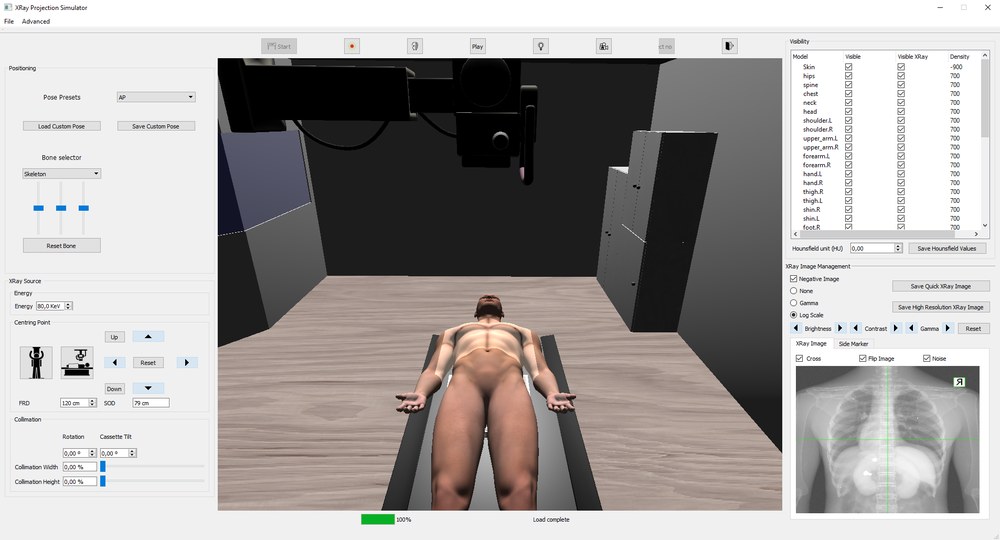
Abstract
gVirtualXray (gVXR) is a programming interface framework to simulate realistic X-ray projections in realtime on graphics processing units (GPUs). It solves the Beer-Lambert law (attenuation law) using a deterministic X-ray simulation algorithm based on 3D computer graphics, namely rasterisation. Implemented as multi-pass rendering makes it more computationally optimal than the ray-tracing technique, which is a brute-force and straightforward approach to simulate X-ray images. Although written in C++ using OpenGL and its shading language (GLSL) to leverage the GPU, gVXR is available for other programming languages such as Python. Extensive validation studies, including comparisons with Monte Carlo simulations and real experimental data, have confirmed the accuracy of gVXR’s simulations. gVXR was initially used in medical virtual reality (VR) for training purposes. It was then used in medical physics, and high-throughput data applications including mathematical optimisation and machine learning (ML). Micro-imaging studies on the C. elegans biological model are also reported.
2023
EuroVis 2023, Leipzig, Germany
First Prize: Visual Exploration, Analysis, and Communication of Physiological Processes
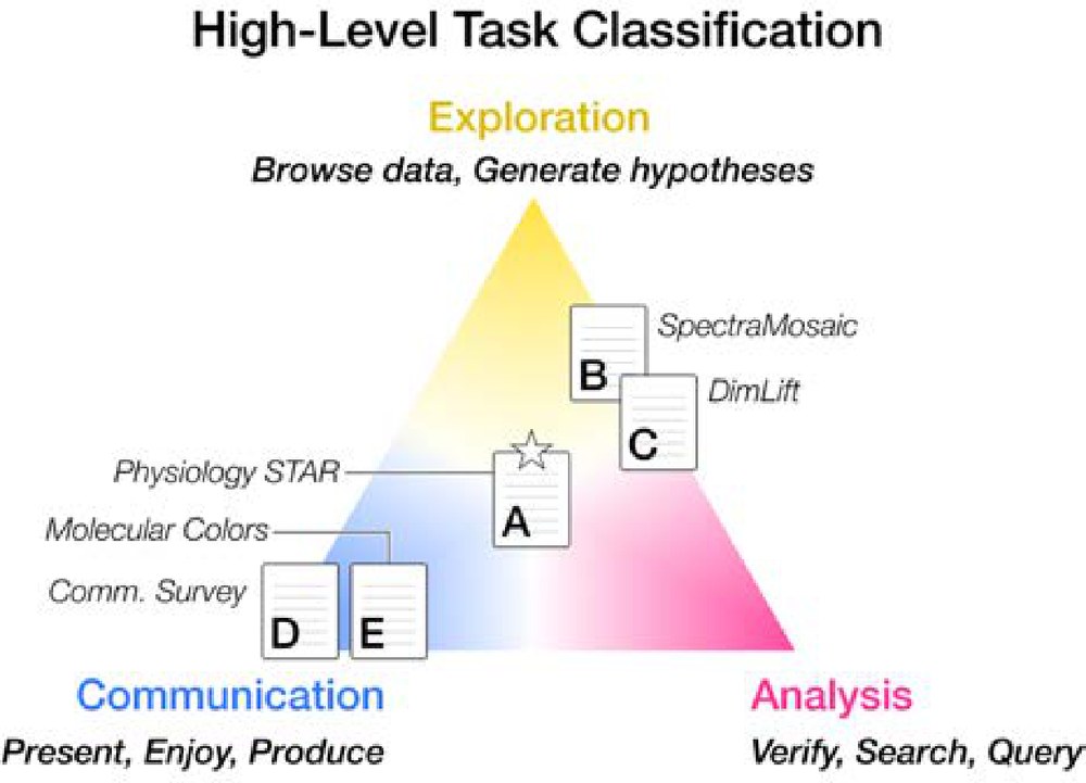
Abstract
Describing the myriad biological processes occurring in living beings over time, the science of physiology is complex and critical to our understanding of how life works. Physiology spans many spatio-temporal scales to combine and bridge from the basic sciences (biology, physics, and chemistry) to medicine. Recent years have seen an explosion of new and finer-grained experimental and acquisition methods to characterize these data. The volume and complexity of these data necessitate effective visualizations to complement standard analysis practice. Visualization approaches must carefully consider and be adaptable to the user’s main task, be it exploratory, analytical, or communication-oriented. This research contributes to the areas of theory, empirical findings, methods, applications, and research replicability in visualizing physiology. Our overarching theme is the cross-disciplinary application of medical illustration and visualization techniques to address challenges in exploring, analyzing, and communicating aspects of human physiology to audiences with differing expertise.
Second Prize: Visualizing Carotid Stenoses for Stroke Treatment and Prevention
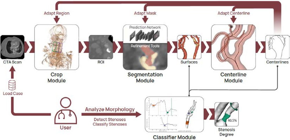
Abstract
Analyzing carotid stenoses – potentially lethal constrictions of the brain-supplying arteries – is a critical task in clinical stroke treatment and prevention. Determining the ideal type of treatment and point for surgical intervention to minimize stroke risk is considerably challenging. We propose a collection of visual exploration tools to advance the assessment of carotid stenoses in clinical applications and research on stenosis formation. We developed methods to analyze the internal blood flow, anatomical context, vessel wall composition, and to automatically and reliably classify stenosis candidates. We do not presume already segmented and extracted surface meshes but integrate streamlined model extraction and pre-processing along with the result visualizations into a single framework. We connect multiple sophisticated processing stages in one user interface, including a neural prediction network for vessel segmentation and automatic global diameter computation. We enable retrospective user control over each processing stage, greatly simplifying error detection and correction. The framework was developed and evaluated in multiple iterative user studies, involving a group of eight specialists working in stroke care (radiologists and neurologists). It is publicly available, along with a database of over 100 carotid bifurcation geometries that were extracted with the framework from computed tomography data. Further, it is a vital part of multiple ongoing studies investigating stenosis pathophysiology, stroke risk, and the necessity for surgical intervention.
Third Prize: Transdisciplinary Visualization of Aortic Dissections
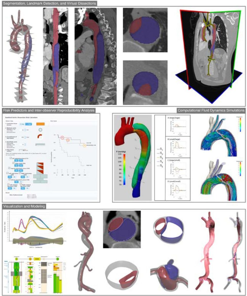
Abstract
Aortic dissection is a life-threatening condition caused by the abrupt formation of a secondary blood flow channel within the vessel wall. Patients surviving the acute phase remain at high risk for late complications, such as aneurysm formation and aortic rupture. The timing of these complications is variable, making long-term imaging surveillance crucial for aortic growth monitoring. Morphological characteristics of the aorta, its hemodynamics, and, ultimately, risk models impact treatment strategies. Providing such a wealth of information demands expertise across a broad spectrum to understand the complex interplay of these influencing factors. We present results of our longstanding transdisciplinary efforts to confront this challenge. Our team has identified four key disciplines, each requiring specific expertise overseen by radiology: lumen segmentation and landmark detection, risk predictors and inter-observer analysis, computational fluid dynamics simulations, and visualization and modeling. In each of these disciplines, visualization supports analysis and serves as communication medium between stakeholders, including patients. For each discipline, we summarize the work performed, the related work, and the results.
2021
EuroVis 2021, Zurich, Switzerland
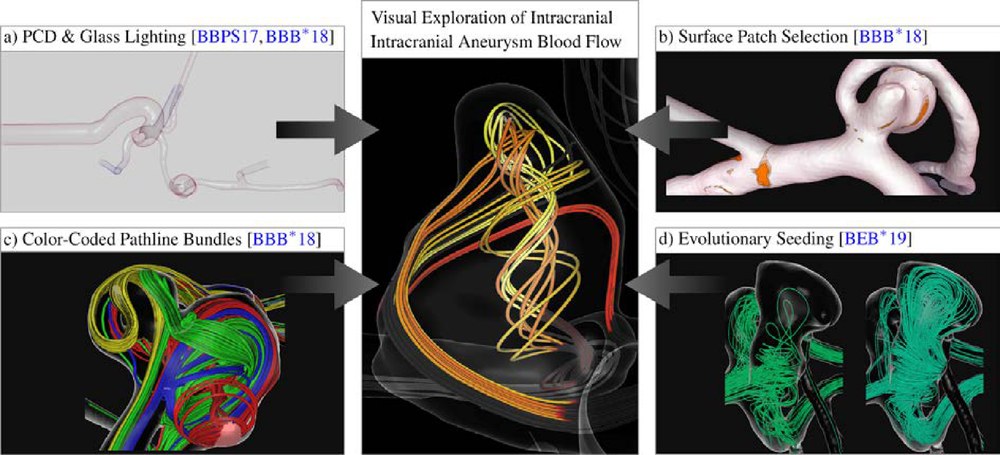
First Prize: Visual Exploration of Intracranial Aneurysm Blood Flow Adapted to the Clinical Researcher
Abstract
Rupture risk assessment is a key to devise patient-specific treatment plans of cerebral aneurysms. To understand and predict the development of aneurysms and other vascular diseases over time, both hemodynamic flow patterns and their effect on the vessel surface need to be analyzed. Flow structures close to the vessel wall often correlate directly with local changes in surface parameters, such as pressure or wall shear stress. However, especially for the identification of specific blood flow characteristics that cause local startling parameters on the vessel surface, like elevated pressure values, an interactive analysis tool is missing. In order to find meaningful structures in the entirety of the flow, the data has to be filtered based on the respective explorative aim. Thus, we present a combination of visualization, filtering and interaction techniques for explorative analysis of blood flow with a focus on the relation of local surface parameters and underlying flow structures. In combination with a filtering-based approach, we propose the usage of evolutionary algorithms to reduce the overhead of computing pathlines that do not contribute to the analysis, while simultaneously reducing the undersampling artifacts. We present clinical cases to demonstrate the benefits of both our filter-based and evolutionary approach and showcase its potential for patient-specific treatment plans.
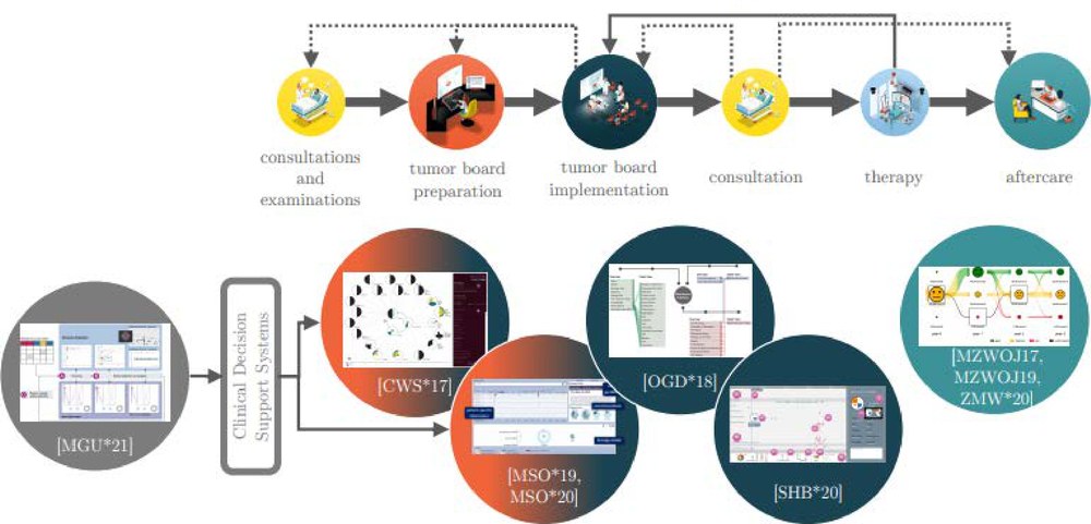
Second Prize: Visual Assistance in Clinical Decision Support
Abstract
Clinical decision-making for complex diseases such as cancer aims at finding the right diagnosis, optimal treatment or best aftercare for a specific patient. The decision-making process is very challenging due to the distributed storage of patient information entities in multiple hospital information systems, the required inclusion of multiple clinical disciplines with their different views of disease and therapy, and the multitude of available medical examinations, therapy options and aftercare strategies. Clinical Decision Support Systems (CDSS) address these difficulties by presenting all relevant information entities in a concise manner and providing a recommendation based on interdisciplinary disease- and patient-specific models of diagnosis and treatment. This work summarizes our research on visual assistance for therapy decision-making. We aim at supporting the preparation and implementation of expert meetings discussing cancer cases (tumor boards) and the aftercare consultation. In very recent work, we started to address the generation of models underlying a CDSS. The developed solutions combine state-of-the-art interactive visualizations with methods from statistics, machine learning and information organization.
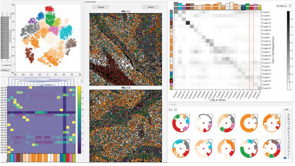
Third Prize: Visual Analysis of Tissue Images at Cellular Level
Abstract
The detailed analysis of tissue composition is crucial for the understanding of tissue functionality. For example, the location of immune cells related to a tumour area is highly correlated with the effectiveness of immunotherapy. Therefore, experts are interested in presence of cells with specific characteristics as well as the spatial patterns they form. Recent advances in single-cell imaging modalities, producing high-dimensional, high-resolution images enable the analysis of both of these features. However, extracting useful insight on tissue functionality from these high-dimensional images poses serious and diverse challenges to data analysis. We have developed an interactive, data-driven pipeline covering the main analysis challenges experts face, from the pre-processing of images via the exploration of tissue samples to the comparison of cohorts of samples. All parts of our pipeline have been developed in close collaboration with domain experts and are already a vital part in their daily analysis routine.
2019
Eurographics 2019, Genova, Italy
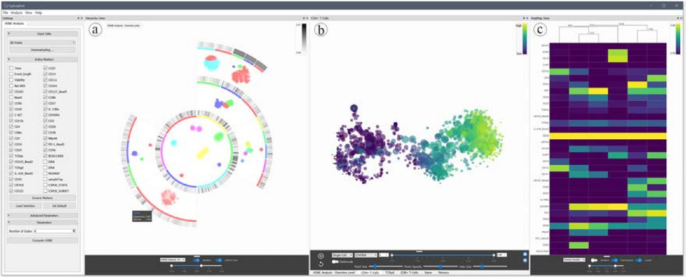
First Prize: Cytosplore: Interactive Visual Single-Cell Profiling of the Immune System
Abstract
Recent advances in single-cell acquisition technology have led to a shift towards single-cell analysis in many fields of biology. In immunology, detailed knowledge of the cellular composition is of interest, as it can be the cause of deregulated immune responses, which cause diseases. Similarly, vaccination is based on triggering proper immune responses; however, many vaccines are ineffective or only work properly in a subset of those who are vaccinated. Identifying differences in the cellular composition of the immune system in such cases can lead to more precise treatment. Cytosplore is an integrated, interactive visual analysis framework for the exploration of large single-cell datasets. We have developed Cytosplore in close collaboration with immunology researchers and several partners use the software in their daily workflow. Cytosplore enables efficient data analysis and has led to several discoveries alongside high-impact publications.
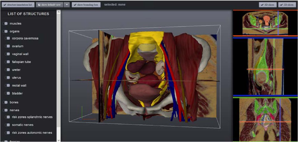
Second Prize: Model-based Visualization for Medical Education and Training
Abstract
Anatomy, or the study of the structure of the human body, is an essential component of medical education. Certain parts of human anatomy are considered to be more complex to understand than others, due to a multitude of closely related structures. Furthermore, there are many potential variations in anatomy, e.g., different topologies of vessels, and knowledge of these variations is critical for many in medical practice. Some aspects of individual anatomy, such as the autonomic nerves, are not visible in individuals through medical imaging techniques or even during surgery, placing these nerves at risk for damage. 3D models and interactive visualization techniques can be used to improve understanding of this complex anatomy, in combination with traditional medical education paradigms. We present a framework incorporating several advanced medical visualization techniques and applications for teaching and training purposes, which is the result of an interdisciplinary project. In contrast to previous approaches which focus on general anatomy visualization or direct visualization of medical imaging data, we employ model-based techniques to represent variational anatomy, as well as anatomy not visible from imaging. Our framework covers the complete spectrum including general anatomy, anatomical variations, and anatomy in individual patients. Applications within our framework were evaluated positively with medical users, and our educational tool for general anatomy is in use in a Massive Open Online Course (MOOC) on anatomy, which had over 17000 participants worldwide in the first run.
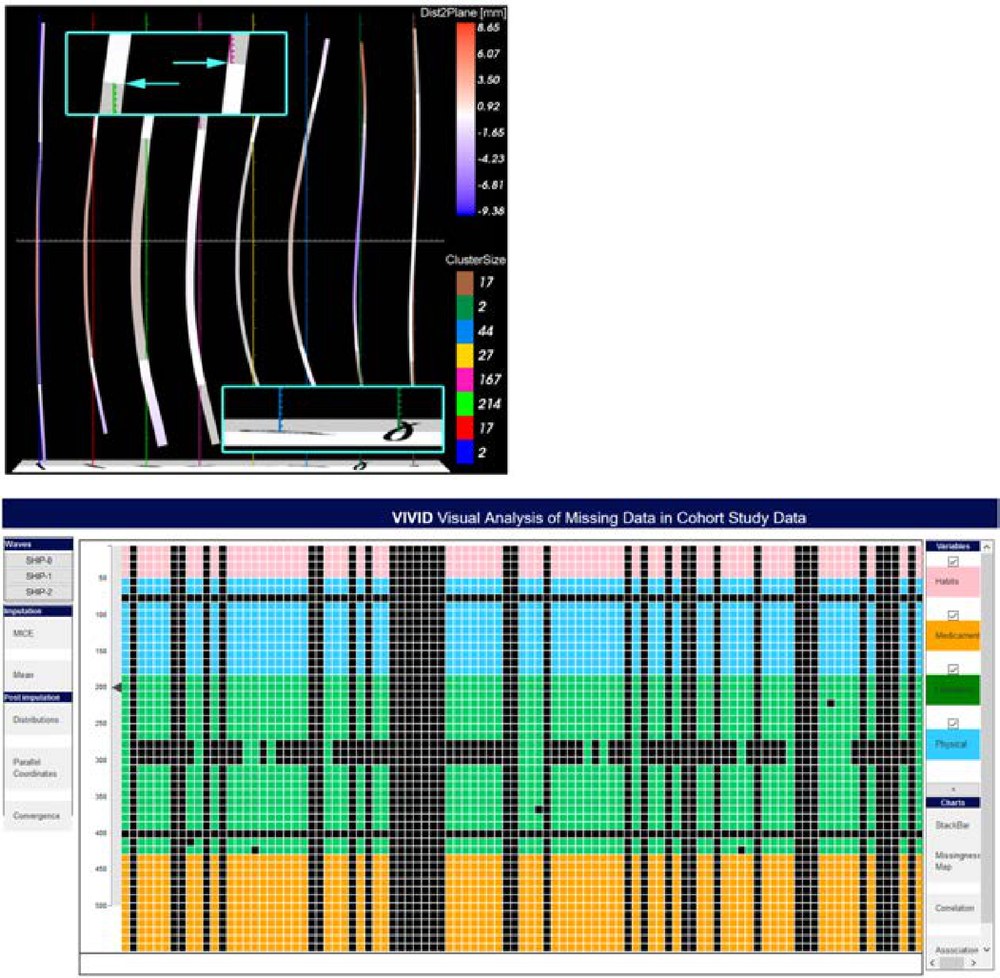
Third Prize: Visual Analytics for Epidemiology
Abstract
We present visual analytics methods to analyze epidemiologic cohort studies. We consider the automatic identification of strong correlations and of subgroups that deviate from the global mean with respect to their risk for health disorders. Moreover, we tackle missing value problems and discuss appropriate imputation strategies and visual analytics support.
2017
Eurographics 2017, Lyon, France
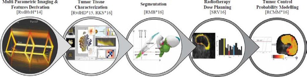
First Prize: Visual Analytics for Digital Radiotherapy: Towards a Comprehensible Pipeline
Abstract
Prostate cancer is one of the most frequently occurring types of cancer in males. It is often treated with radiation therapy, which aims at irradiating tumors with a high dose, while sparing the surrounding healthy tissues. In the course of the years, radiotherapy technology has undergone great advancements. However, tumors are not only different from each other, they are also highly heterogeneous within, consisting of regions with distinct tissue characteristics, which should be treated with different radiation doses. Tailoring radiotherapy planning to the specific needs and intra-tumor tissue characteristics of each patient is expected to lead to more effective treatment strategies. Currently, clinical research is moving towards this direction, but an understanding of the specific tumor characteristics of each patient, and the integration of all available knowledge into a personalizable radiotherapy planning pipeline are still required. The present work describes solutions from the field of Visual Analytics, which aim at incorporating the information from the distinct steps of the personalizable radiotherapy planning pipeline, along with eventual sources of uncertainty, into comprehensible visualizations. All proposed solutions are meant to increase the – up to now, limited – understanding and exploratory capabilities of clinical researchers. These approaches contribute towards the interactive exploration, visual analysis and understanding of the involved data and processes at different steps of the radiotherapy planning pipeline, creating a fertile ground for future research in radiotherapy planning.
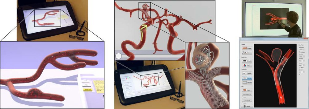
Second Prize: Sketching and Annotating Vascular Structures to Support Medical Teaching, Treatment Planning and Patient Education
Abstract
In clinical practice, hand drawn sketches are employed to express concepts and are an efficient method for the discussion of complex issues. We present computer graphic methods to improve and support the creation and annotation of complex sketches, resulting in a more clear, expressive and understandable result. For this, we consider the medical areas of teaching, treatment planning and patient education. Our applications allow students, educators, physicians and patients to sketch and annotate vascular structures, their pathologies and treatment options as well as to simulate and illustrate blood flow. The used sketching approaches take advantage of semi-immersive environments as well as interactive whiteboards to enable the creation of vessels either in their spatially complex 3D representation or as a simplified 2D illustration. We evaluate our work in interviews with physicians and user studies to assess their usability and to reveal their benefits to support the respective medical domain.
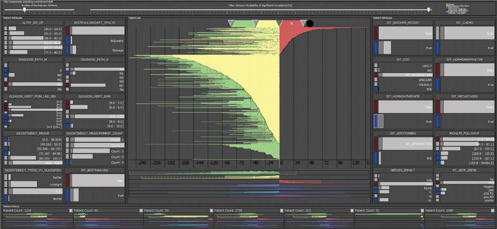
Third Prize: Visual Computing for Big Data Analysis in Prostate Cancer Research
Abstract
Data-centered research is becoming increasingly important in prostate cancer research where a long-term goal is a sound prognosis prior to surgery. We have developed a visual computing technology that contributes to this paradigm change in clinical research and practice for electronic health records (EHR) in this area. This visual-interactive system, developed in close collaboration with medical researchers, helps clinicians efficiently and effectively visualize single and multiple patient histories at a glance, create cohorts of patients for clinical tests, as well as generate and validate hypotheses.
2015
Eurographics 2015, Zurich, Switzerland
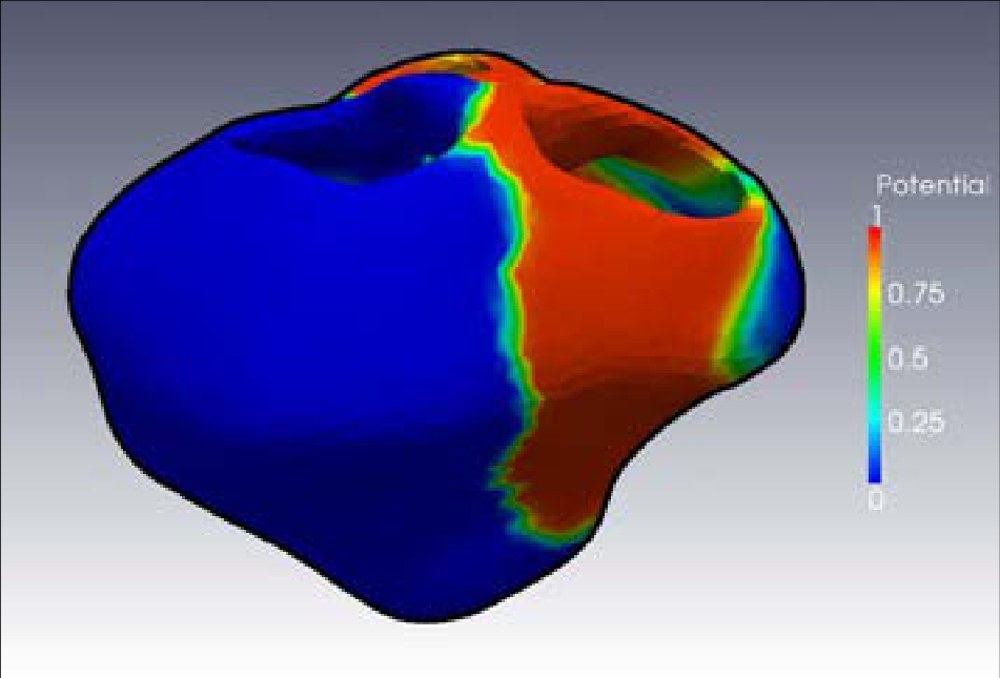
First Prize: Surgery Training, Planning and Guidance Using the SOFA Framework
Abstract
In recent years, an active development of novel technologies dealing with medical training, planning and guidance has become an increasingly important area of interest in both research and health-care manufacturing. A combination of advanced physical models, realistic human-computer interaction and growing computational power is bringing new solutions in order to help both medical students and experts to achieve a higher degree of accuracy and reliability in surgical interventions. In this paper, we present three different examples of medical physically-based simulations implemented in a common software platform called SOFA. Each example represents a different application: training for cardiac electrophysiology, pre-operative planning of cryosurgery and per-operative guidance for laparoscopy. The goal of this presentation is to evaluate the realism, accuracy and efficiency of the simulations, as well as to demonstrate the potential and flexibility of the SOFA platform.
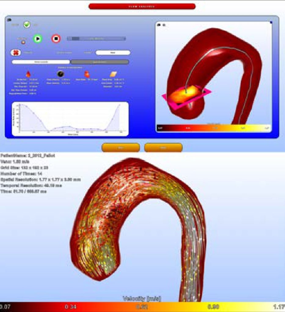
Second Prize: Guided Analysis of Cardiac 4D PC-MRI Blood Flow Data
Abstract
Four-dimensional phase-contrast magnetic resonance imaging (4D PC-MRI) allows the non-invasive acquisition of temporally resolved, three-dimensional blood flow information. Quantitative and qualitative data analysis helps to assess the cardiac function, severity of diseases and find indications of different cardiovascular pathologies. However, various steps are necessary to achieve expressive visualizations and reliable results. This comprises the correction of special MR-related artifacts, the segmentation of vessels, flow integration with feature extraction and the robust quantification of clinically important measures. A fast and easy-to-use processing pipeline is essential since the target user group are physicians. We present a system that offers such a guided workflow for cardiac 4D PC-MRI data. The aorta and pulmonary artery can be analyzed within ten minutes including vortex extraction and robust determination of the stroke volume as well as the percentaged backflow. 64 datasets of healthy volunteers and of patients with variable diseases such as aneurysms, coarctations and insufficiencies were processed so far.
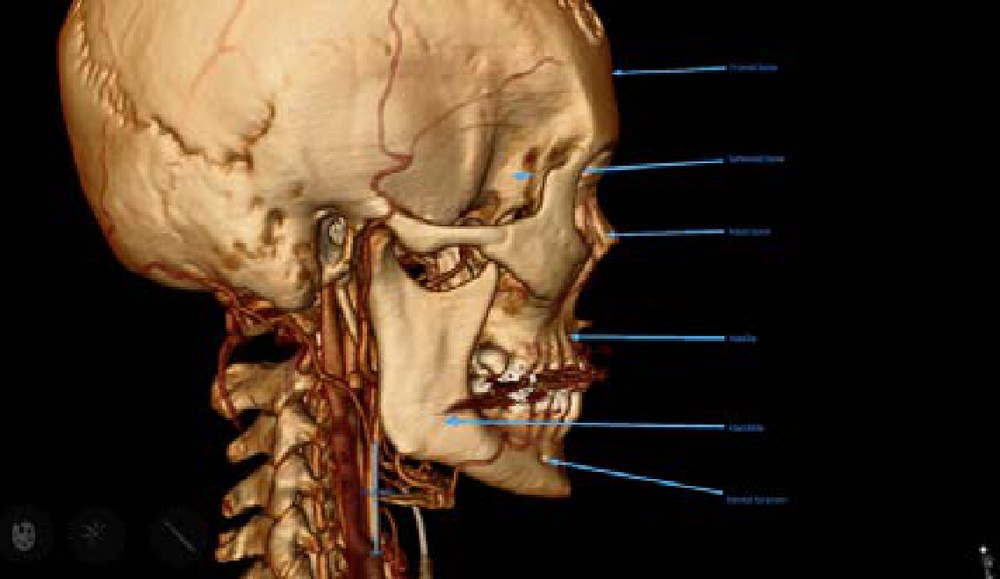
Third Prize: Multi-Touch Table System for Medical Visualization
Abstract
Medical imaging plays a central role in a vast range of healthcare practices. While the usefulness of 3D visualizations is well known, the adoption of such technology has previously been limited in many medical areas. This paper, awarded the Dirk Bartz Prize for Visual Computing in Medicine 2015, describes the development of a medical multi-touch visualization table that successfully has reached its aim to bring 3D visualization to a wider clinical audience. The descriptions summarize the targeted clinical scenarios, the key characteristics of the system, and the user feedback obtained.
2013
Eurographics 2013, Girona, Spain
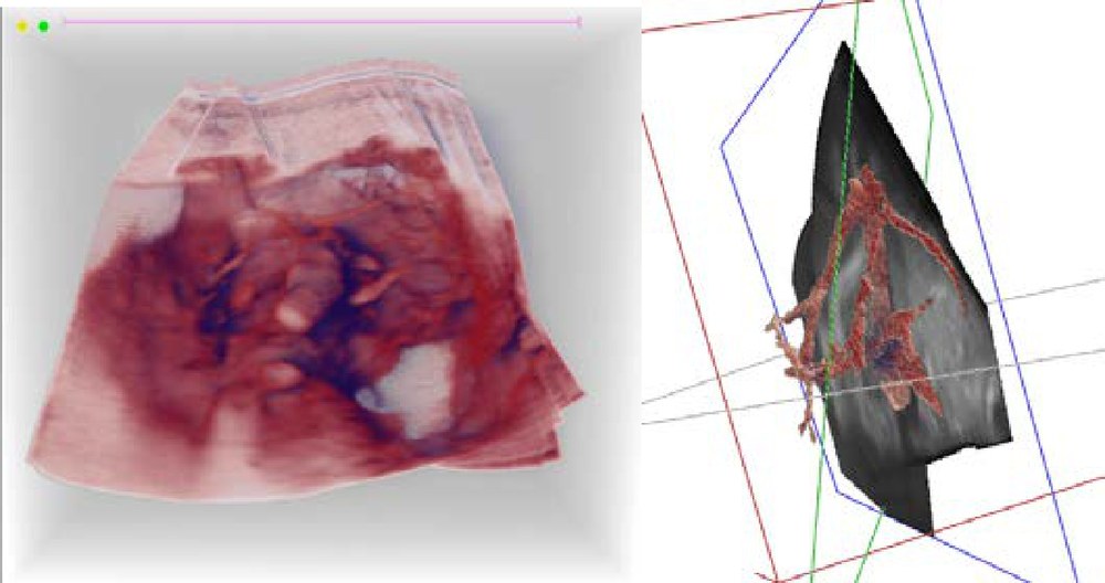
First Prize: High-Quality 3D Visualization of In-Situ Ultrasonography
Abstract
In recent years medical ultrasound has experienced a rapid development in the quality of real-time 3D ultrasound (US) imaging. The image quality of the 3D volume that was previously possible to achieve within the range of a few seconds, is now possible in a fraction of a second. This technological advance offers entirely new opportunities for the use of US in the clinic. In our project, we investigate how real-time 3D US can be combined with high-performance processing of today’s graphics hardware to allow for high-quality 3D visualization and precise navigation during the examination.
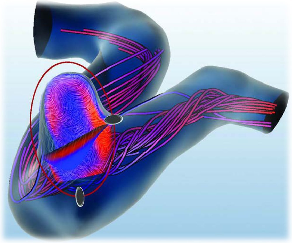
Second Prize: Effective Visual Exploration of Hemodynamics in Cerebral Aneurysms
Abstract
Cerebral aneurysms are a pathological vessel dilatation that bear a high risk of rupture. For the understanding of this risk, the analysis of hemodynamic information plays an important role in clinical research. These information are obtained by computational fluid dynamics (CFD) simulations. Thus, an effective visual exploration of patient-specific blood flow behavior in cerebral aneurysms was developed to support the domain experts in their investigation process. We present advanced visualization and interaction techniques, which provide an overview, focus-and-context views as well as multi-level explorations. Moreover, an automatic extraction process of qualitative flow characteristics, which are correlated with the risk of rupture is introduced. Although not established in clinical routine yet, interviews and informal user studies confirm the usefulness of these methods.
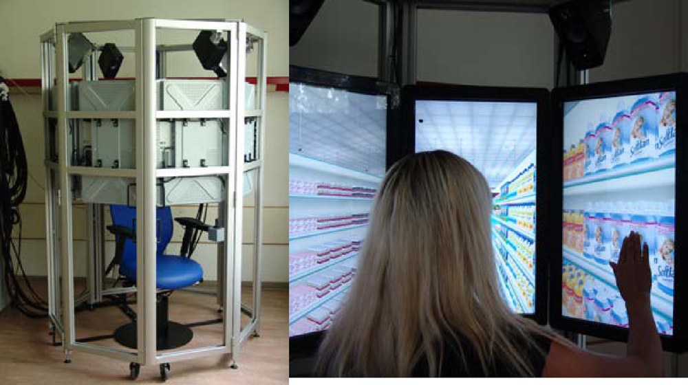
Third Prize: OctaVis: A Virtual Reality System for Clinical Studies and Rehabilitation
Abstract
Brain function disorders, resulting for instance from stroke, epilepsy, or other incidents can be partially recovered by rehabilitation training. Performing neuro-rehabilitation in virtual reality systems allows for training scenarios close to daily tasks, is easily adaptable to the patients’ needs, is fully controllable by clinical staff, and guarantees patient safety at all times. In this paper, we describe the OCTAVIS system, a novel virtual reality platform developed primary for clinical studies with and rehabilitation training of patients with brain function disorders. To meet the special requirements for clinical use, our system has been designed with ease of use, ease of maintenance, patient safety, space and cost efficiency in mind. Our system has been successfully deployed to four hospitals, where it is used for rehabilitation training and clinical studies. We report first results of these studies, demonstrating that our system is immersive, easy to use, and supportive for rehabilitation purposes.
2011
Eurographics 2011, Llandudno, United Kingdom
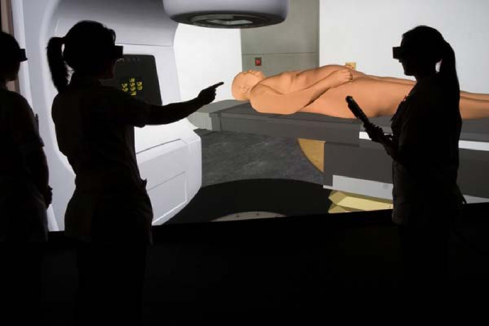
Shared First Prize: A Virtual Environment for Radiotherapy Training and Education – VERT
Abstract
A report in 2007 to the UK Government identified a crisis in England for training staff and students for the radiotherapy treatment of cancer. The Hull authors have developed an immersive life size virtual environment of a radiotherapy treatment room, known as VERT, to address this problem. VERT provides the trainee with models, simulation, enhanced visualization and training aids for treatment of virtual patients in a virtual treatment room. In 2007 VERT systems for radiotherapy training were established for training purposes at the University Aarhus Hospital (Denmark), Birmingham City University (UK) and the University of Ulster (UK). There are now some 70 VERT systems around the world. This paper reports on the simulation and visualization capabilities and reports on the use of VERT from the Aarhus University hospital and on the national evaluation of VERT in the UK [AC10]. These reports clearly indicate the clinical benefit of using a virtual environment approach, such as VERT, for training and education in radiotherapy.
Shared First Prize: The Tumor Therapy Manager and its Clinical Impact
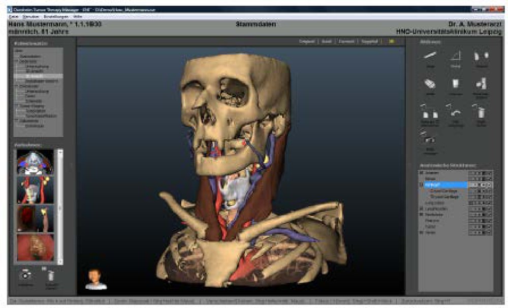
Abstract
Visual exploration of CT and MRI datasets in clinical practice is still dominated by slice-based viewing. Volume rendering is now widely available but seen primarily as a tool for a fast overview, and only rarely as a visualization to directly support clinical decisions. Research projects aiming at advanced 3D visualizations, such as smart visibility and illustrative renderings, usually fail to meet clinical demands, since the visualizations are not dedicated to specific diagnostic or treatment planning questions. Moreover, they are unfamiliar to users who need reliable and familiar visualizations as a basis for their crucial decisions. Discussions with clinical practicians reveal that parameterization of visual effects is too cumbersome and resulting visualizations are often too complex. We describe and discuss long-term experiences on developing, testing, and refining image analysis and visualization techniques for ENT surgery planning based on CT data. While visual quality and a faithful rendition of spatial relations indeed are essential, it turned out to be superior to generate sequences of rather simple 3D visualizations directly supporting specific treatment questions instead of presenting many anatomic structures simultaneously. We report on the actual clinical use of the system and discuss how it changed the surgical planning workflow.
Third Prize: AVM-Explorer: Multi-Volume Visualization of Vascular Structures for Planning of Cerebral AVM Surgery
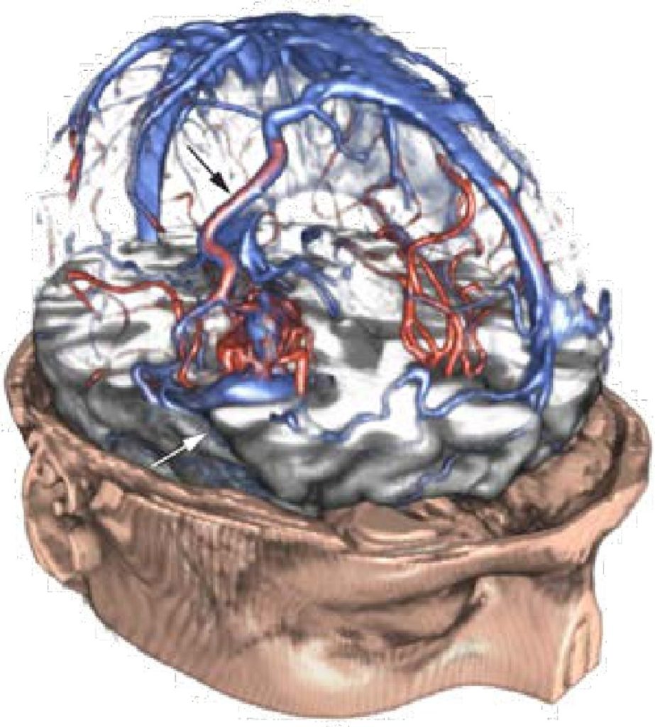
Abstract
Arteriovenous malformations (AVMs) of the brain are rare vascular disorders characterized by the presence of direct connections between cerebral arteries and veins. Preoperative planning of AVM surgery is a challenging task. The neurosurgeon needs to gain a detailed understanding of both the pathoanatomy of the lesion as well as its location and spatial relation to critical functional areas and white matter fiber bundles at risk. A crucial element during this planning phase is the precise identification of feeding arteries, draining veins, and arteries en passage”. To this end, a variety of imaging modalities for displaying neurovascular structures exists, both tomographic as well as projection based. However, the conventional 2D slice based review of such data is not well suited to help understanding the complex angioarchitecture of an AVM. In this paper, we demonstrate how stateof- the-art techniques from the fields of computer graphics and image processing can support neurosurgeons with the challenge of creating a mental 3D model of the lesion and understanding its internal structure. To evaluate the clinical value of our method, we present results from three case studies along with the medical assessment of an experienced neurosurgeon.
Honorable Mention: Interactive Visualization Techniques for Neurosurgery Planning
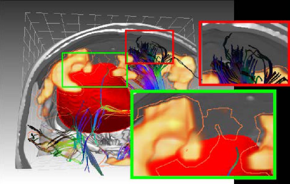
Abstract
We present concepts for pre-operative planning of brain tumor resections. The proposed system uses a combination of traditional and novel visualization techniques rendered in real-time on modern GPUs in order to support neurosurgeons during intervention planning. A set of multimodal 2D and 3D renderings conveys the relation between the lesion and the various structures at risk and also depicts data uncertainty. To facilitate efficient interactions while providing a comprehensible visualization, all employed views are linked. Furthermore, the system allows the surgeon to interactively define the access path by clicking in the 3D views as well as to perform distance measurements in 2D and 3D.
Honorable Mention: FEMONUM: A Framework for Whole Body Pregnant Woman Modeling from Ante-Natal Imaging Data
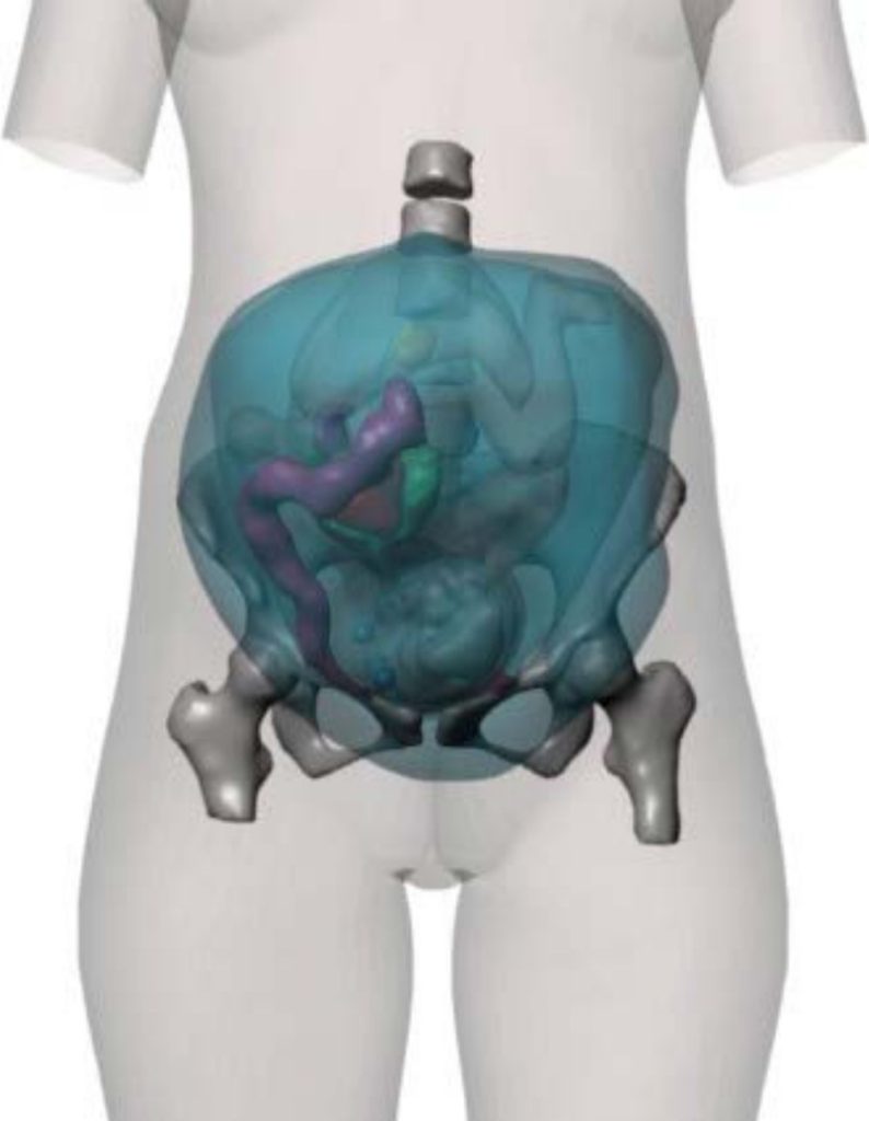
Abstract
Anatomical models of pregnant women can be used in several applications such as numerical dosimetry to assess the potential effects of electromagnetic fields on biological tissues, or medical simulations for delivery planning. Recent advances in medical imaging have enabled the generation of realistic and detailed models of human beings. This paper describes FEMONUM, a complete methodological framework for the construction of pregnant woman models based on medical images and their segmentation. FEMONUM combines several computer graphics methods, such as surface reconstruction and physics-based computer animation to model and deform pregnant women abdomens, to simulate different fetal positions and sizes and also different morphologies of the mother, represented with a synthetic woman body envelope. A set of 16 models, anatomically validated by clinical experts, is presented and is made available online to the scientific community. These models include detailed information on the utero-fetal units and cover different gestational stages with various fetal positions.
2009
Eurographics 2009, Munich, Germany
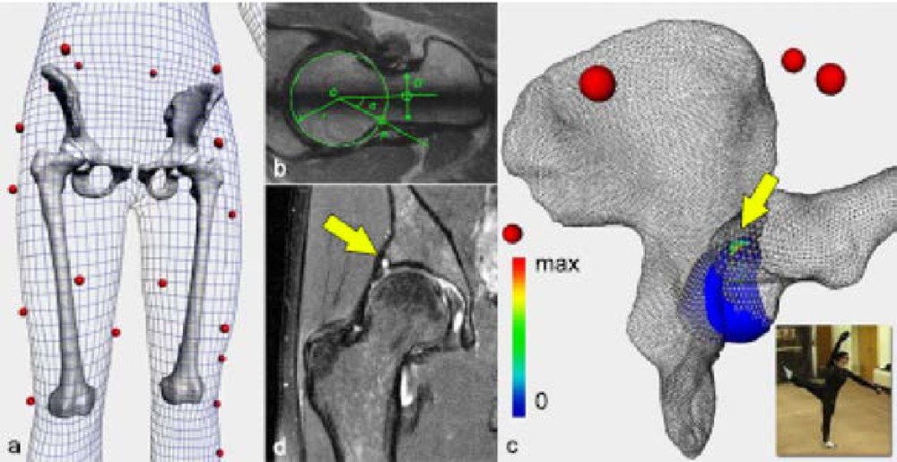
First Prize: Virtual Hip Joint: from Computer Graphics to Computer-Assisted Diagnosis
Abstract
Osteoarthritis (OA) is a major musculoskeletal disorder which causes are not always fully understood. Femoroacetabular impingements such as cam/ pincer cannot always explain observed OA in hips with normal morphology. This paper investigates the hypothesis of extreme repetitive movements as a source of cartilage degeneration. We present a clinical study conducted with professional ballet dancers and a methodology to perform functional simulations of the hip joint in extreme postures. Throughout the process, various computer graphics techniques are used, like motion capture, 3D body scanning and physically-based models. In addition to accelerate and strengthen some tasks, these techniques strongly participate in the clinical understanding of OA related to motion. Preliminary results have indeed shown a significant correlation between the location of impingements and radiologically observed damage zones in the labrum cartilage.
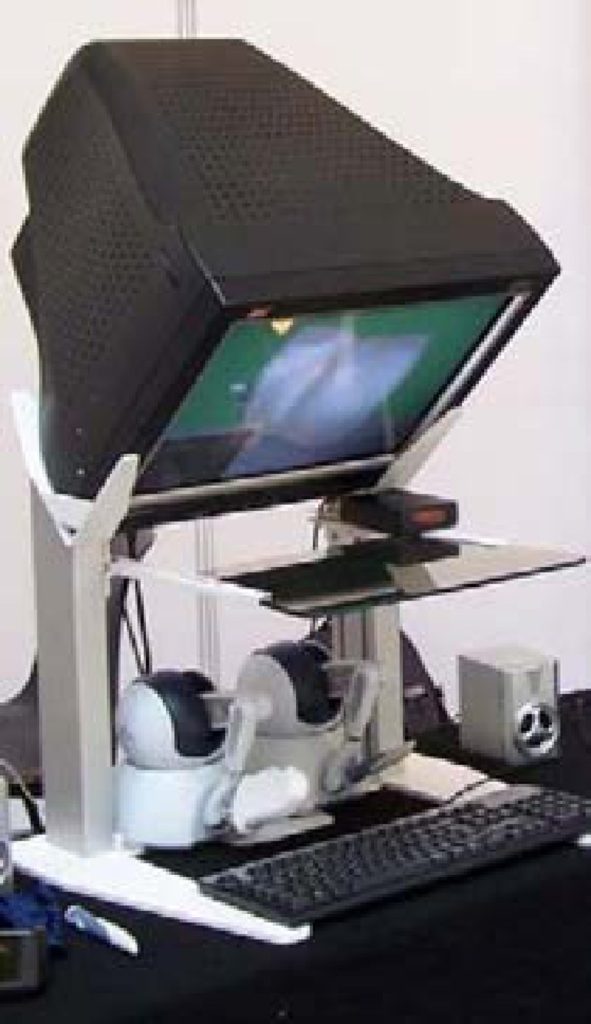
Second Prize: ImaGINe-S: Imaging Guided Interventional Needle Simulation
Abstract
We present an integrated system for training visceral needle puncture procedures. Our aim is to provide a cost effective and validated training tool that uses actual patient data to enable interventional radiology trainees to learn how to carry out image-guided needle puncture. The input data required is a computed tomography scan of the patient that is used to create the patient specific models. Force measurements have been made on real tissue and the resulting data is incorporated into the simulator. Respiration and soft tissue deformations are also carried out to further improve the fidelity of the simulator.
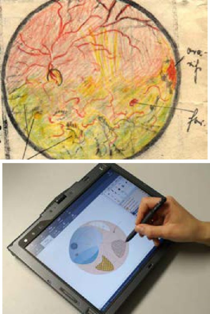
Shared Third Prize: GREiF – Graphical Documentation of Retinal Findings Using a Standardized Digital Symbol Library
Abstract
Aim: To develop a software package that improves standardized clinical documentation of retinal findings. In clinical routine retinal findings are usually documented with sketchy free-hand drawings and supplementary handwritten remarks. Documentation features of common ophthalmologic software products include only simple sketching functions, which are limited to change location, size or colour of graphical primitives (e. g. ovals, rectangles, lines, textboxes . . .). As a result a feasible creation of standardized graphical documentations in retinal imaging is almost impossible. Methods: We developed a java-based software tool that features quick and intuitive generation of fundus schemes, which can be printed as findings sheet or digitally archived. Particularly for clinical ophthalmologists we created a set of standardized symbols, which can be digitally rendered for graphical documentation. All symbols were integrated into a graphics library and separated in specific categories: “Preoperative”, “Postoperative”, “Angiomas and tumours”, “Retinopathy of the premature”. The required symbol can be chosen from the library and is simply modelled on the retina scheme by placing anchor points with mouse clicks. Results: Practicability of existing features for graphical documentation of retinal findings is not sufficient, because free-hand drawings are too time-consuming and besides share the risk of false interpretation due to individual handwritings. In contrast to free-hand sketching our software tool not only applies a faster way of graphical creation but additionally improves medical documentation using a standardized symbol library, which also is specifically categorized. Conclusions: Graphical symbols for retinal documentation have found universal acceptance in ophthalmologists for a long time but still the practical use is not efficient in clinical routine. This report shows how the adequate use of software technology can contribute to documentation quality and clinical practice.
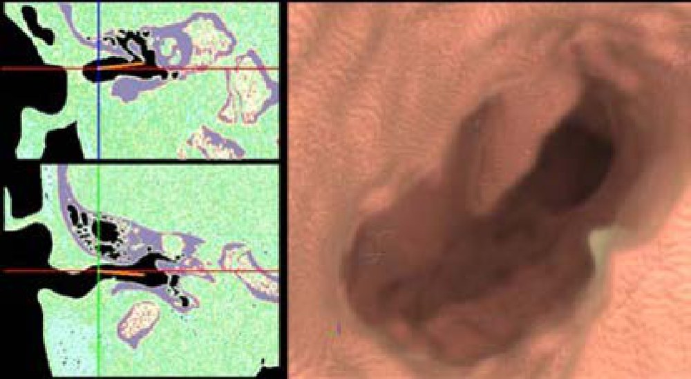
Shared Third Prize: Advanced GPU Volume Rendering for Virtual Endoscopy
Abstract
For difficult cases in endoscopic sinus surgery, a careful planning of the intervention is necessary. Virtual endoscopy enables the visualization of the operating field and additional information, such as risk structures and target structures to be removed. The Sinus Endoscopy system provides the functional range of a virtual endoscopic system with special focus on a realistic representation. Furthermore, by using direct volume rendering, we avoid time-consuming segmentation steps for the use of individual patient datasets. However, the image quality of the endoscopic view can be adjusted in a way that a standard computer with a modern standard graphics card achieves interactive frame rates with low CPU utilization. Thereby, characteristics of the endoscopic view are systematically used for the optimization of the volume rendering speed. As a small standalone application it can be instantly used for surgical planning and patient education. The system was used for preoperative planning in 102 cases, provides useful information for intervention planning (e.g., anatomic variations of the Rec. Frontalis), and closely resembles the intraoperative situation.
2007
Eurographics 2007, Prague, Czech Republic
First Prize: State-of-the-Art Computer Graphics in Neurosurgical Planning and Risk Assessment
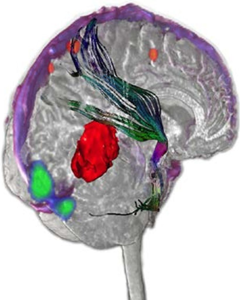
Abstract
We present a novel software assistant that unlocks new potentials in neurosurgical planning and risk assessment. It allows surgeons to approach the task in an intuitive manner, by providing them with the possibility to simultaneously observe all relevant data of a case in synchronized 2D and 3D views. State-of-the-art technologies from the field of computer graphics are combined to allow simultaneous interactive rendering of anatomical and functional MR data in combination with manually segmented objects and slice-based overlays. This allows surgeons to perceive a clearer impression of the anatomical and functional structures affected by an intervention, and especially the way they are related to each other. Thus, it significantly facilitates the finding of an optimal intervention strategy.
Second Prize: A Pen-based Interface for Generating Graphical Reports of Findings in Cardiac Catheterization
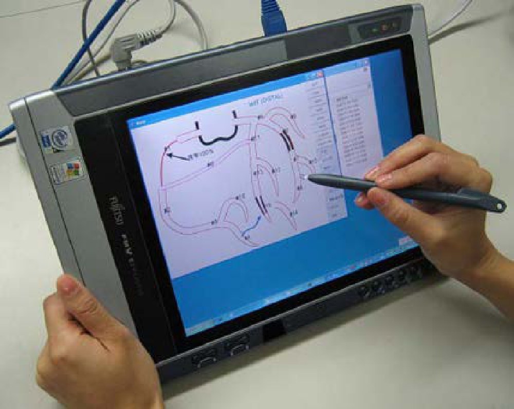
Abstract
This paper introduces a pen-based interface for the graphical reporting of findings in cardiac catheterization. The user can interactively draw, erase, move, and deform coronary arteries as well as record stenoses on them. The lo-cation and degree of each stenosis is represented visually and the doctor can record various treatments such as by-passes and stents on the diagram. In addition, the system automatically extracts semantic information from the graphical representation and stores it in XML format. The system can also generate a table in the format specified by the American Heart Association. This system is useful not only as a tool for efficiently generating reports of findings but also as an effective explanation tool for patients.
Third Prize: Analysis of the Pulmonary Vein Ostia using Cardiac 4DCT for Radiosurgical Ablation
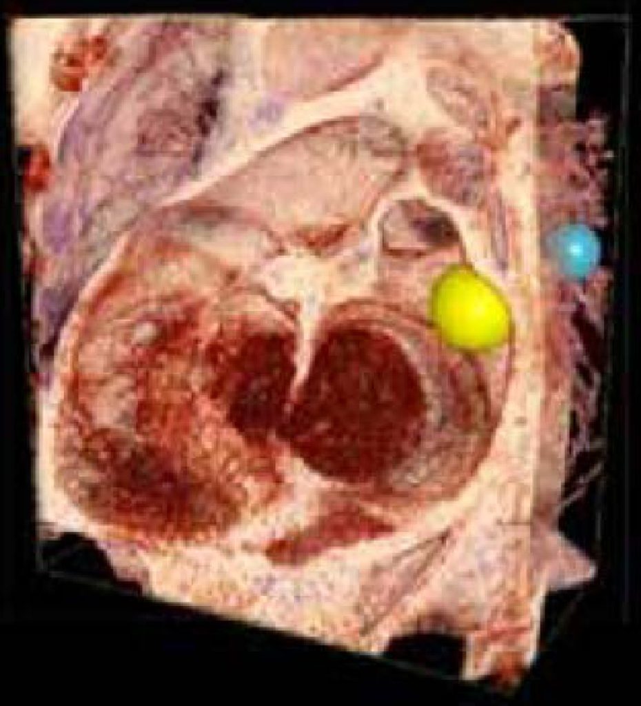
Abstract
A software tool to analyze 4D cardiac CT data sets for planning radiosurgical ablations in the heart is presented. Volume rendering and data processing are performed using a GPU. The user visualizes the data from inside the left atrium and defines the target in 3D using an intuitive user interface. A haptic input device lets the user measure motion at the ostia of the pulmonary veins for radiosurgical treatment planning. This tool has been used effectively for generating radiation treatment plans for animal studies.
2005
Eurographics 2005, Dublin, Ireland
First Prize: A Virtual Reality Toolkit for the Diagnosis and Monitoring of Myocardial Infarctions
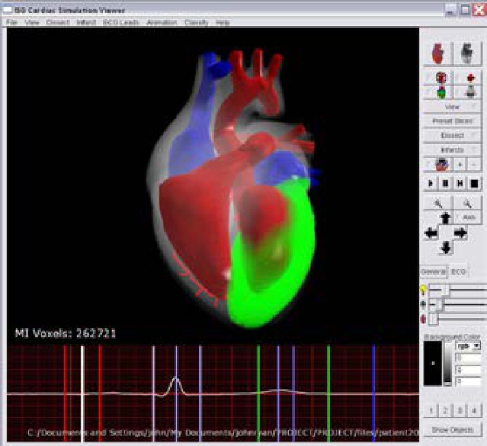
Abstract
We have developed a software system that takes standard electrocardiogram (ECG) input and interprets this input along with user-defined and automatically defined markers to diagnose myocardial infarctions (MI). These pathologies are then automatically represented within a volumetric model of the heart. Over a period of six months 30 patients were monitored using a digital ECG system and this information was used to test and develop our system. It was found that the STEMIs (ST segment Elevation MI) were successfully diagnosed, however NSTEMIs (Non-STEMI), although correctly interpreted, were more ambiguous due to the fact that T wave inversions are sometimes seen on normal ECGs. Control ECGs of normal hearts were also taken. The system correctly interpreted this data as being normal. A standard voxel-count metric was developed so that future work in MI monitoring will be possible. The toolkit was found to be beneficial for three possible uses, as a diagnostic tool for clinicians, as a teaching tool for students and also as an information tool for the patient.
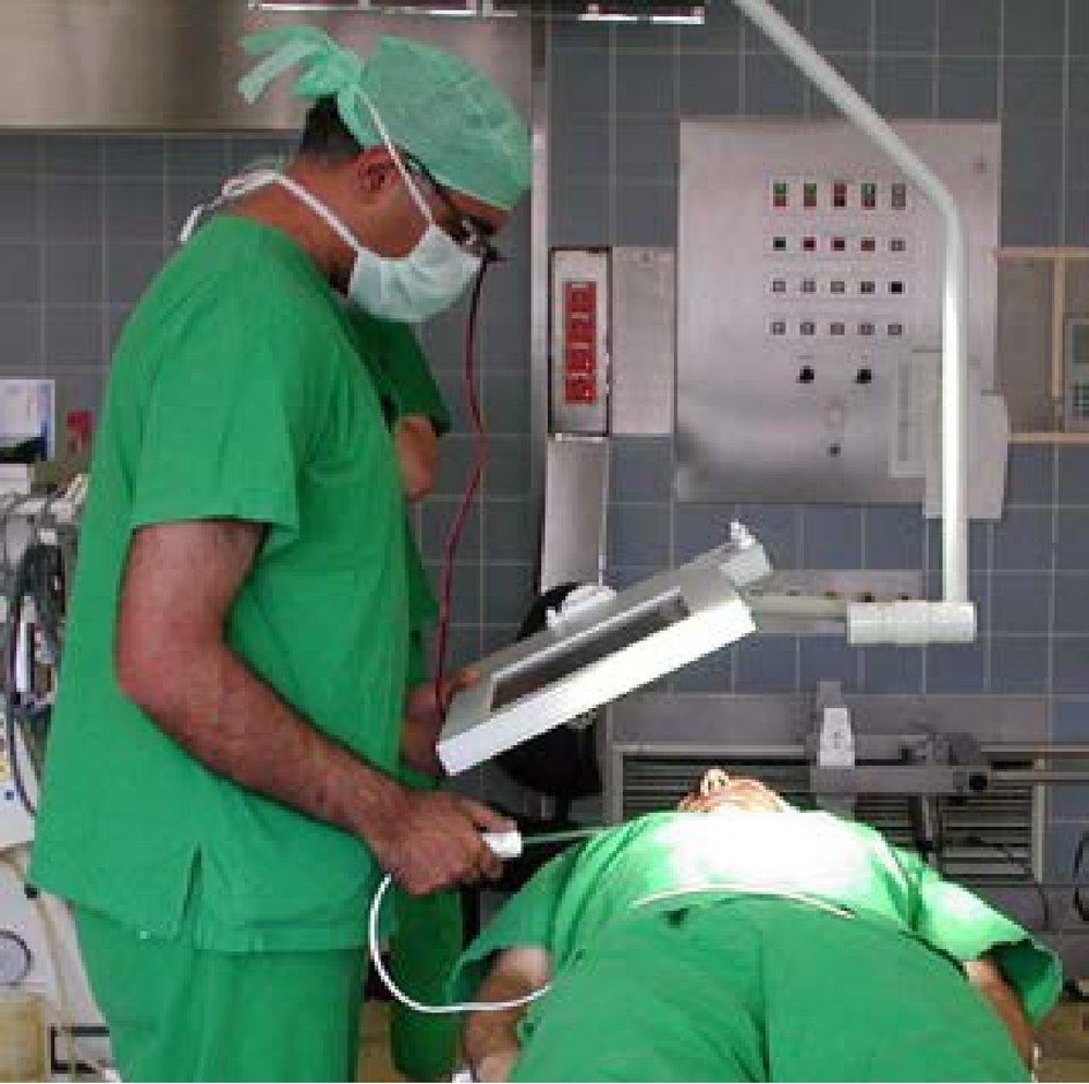
Second Prize: MEDARPA – An Augmented Reality System for Supporting Minimally Invasive Interventions
Info
Fraunhofer IGD, Darmstadt; Computer Graphics Center (ZGDV), Darmstadt; MedCom GmbH, Darmstadt, Germany. No record in the Eurographics Digital Library.
Third Prize: The AngioVis ToolBox